Home
Common Techniques
Classroom Experiments
Virtual Experiments
Tutorials
Games
Glossary
Links
Publishing
Opportunities
About This Site
Contact Us
ZFIN
Cite Us
How to make/use tools for handling zebrafish
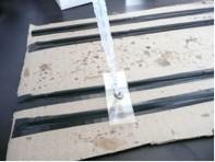
How to Make Bridged Cover Slips
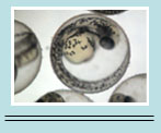
When embryos are viewed by microscopy, it is often useful to mount the embryos in bridged covers slips. These make it possible to roll the embryo into different positions and image them from different views. This is typically done for fixed, stained, embryos. Follow the steps below:
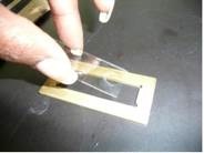
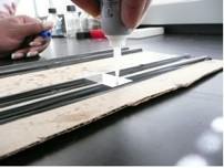
1. Place a clean cover slide onto an elevated surface.
2. Place a couple of droplets of superglue onto the two sides of the cover slide.
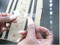
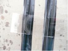
3. Place a cover slip over the superglue on each side of the slide. (You may stack as many as up to 3 cover slips on each side depending on how thick the object is that you are observing. Just superglue the cover slips to each other.)
4. Allow the slide to dry.
If mounting in glycerol:
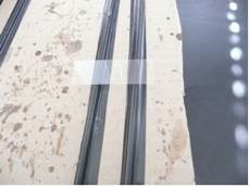
5. Pipette a droplet of glycerol in the middle of the slide.
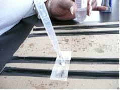
6. Place the specimen in the glycerol. (If the specimen is already in the glycerol, just pipette the both of them together.)
|
|

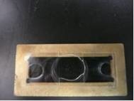
7. Place the slide on a brass ring (download directions for making brass rings).
|
|