
Home
Common Techniques
Classroom Experiments
Virtual Experiments
Tutorials
Games
Glossary
Links
Publishing
Opportunities
About This Site
Contact Us
ZFIN
Cite Us
Antibody Staining
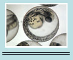
Antibody staining is commonly used in laboratories to locate specific cellular components or molecules on a specimen. It usually occurs in a two-step method, where the primary antibody is used to target the wanted molecule, and the secondary antibody has a fluorescent tag to locate the primary antibody. This process occurs over a period of three days. Embryos are typically inside of Eppendorf tubes (1.5 mL) for all of the following steps.
Day 1
For fish stored in methanol: Re-hydrate the embryos at room temperature with three successive, 5-minute washes, composed of:
1.) 75% methanol/25% PBS
2.) 50% methanol/50% PBS
3.) 25% methanol/75% PBS
Then, rinse with 1X PBS + 0.5% Triton-X100 (250 µL Triton-X100 in 50 mL PBS) for 5 minutes at room temperature. Repeat this step three times while rocking the samples during each wash.
For fish older than 2 days: Incubate in 0.1% collagenase/PBS (25 microliters/10 embryos) for 90 minutes at room temperature. Do not rock the tubes.
Quickly wash twice with 1X PBS + 0.05% Triton-X.
Incubate in PBS/BSA/DMSO/Triton-X100 (PBDT: bring 1 g BSA to 100 mL of PBS, add 1 mL DMSO, and 100 µL Triton-X100) for 1 hr at room temperature. Then rock the tubes.
Incubate in the correct dilution of primary antibody in PBDT at 4.0 C overnight, keeping the tubes rocking.
Day 2
Wash with 1X PBS + 0.05% Triton-X100 for 1 hour at 4.0 C. Wash a total of 4 times, 1 hour each.
Incubate embryos in 1 mL of diluted secondary antibody in PBDT at 4.0 C, rocking overnight. Cover with aluminum foil (assuming you are using a fluorescent secondary that is subject to photobleaching-if not, you can ignore aluminum foil wraps throughout).
Day 3
Wash with PBS for 1 hr at 4.0 C, rocking, 3 times. Keep covered with aluminum foil.
Store in 50% glycerol at 4. 0 C, covered with aluminum foil.